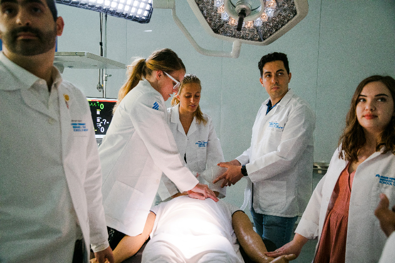
Kaiser Permanente Bernard J. Tyson School of Medicine’s Simulation Center provides hands-on experience in a supportive environment.
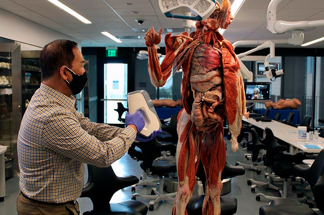
Frank Thai, principal biomedical science technologies consultant for the school, creates a 3D scan of a preserved cadaver. Because 3D cadavers last many years, they’re available to students throughout their medical school experience.
A modern approach to teaching anatomy
Kaiser Permanente Bernard J. Tyson School of Medicine uses cutting-edge technology to teach about the body
When the COVID-19 pandemic emerged, medical schools had to quickly reimagine how to train their students.
Much instruction that would normally happen in person needed to be done virtually due to stay-at-home orders and physical distancing guidelines. Teaching anatomy was especially challenging because students traditionally learn about the structure of the human body by dissecting human cadavers, or bodies, in a lab.
The newly opened Kaiser Permanente Bernard J. Tyson School of Medicine was able to quickly pivot thanks to the school’s first-of-its-kind Anatomy Resource Center. Equipped with both hands-on and virtual tools, the center offers students engaging ways to explore anatomy whether they are on site or learning remotely.
Innovative ways to learn about the body
When students visit the Anatomy Resource Center, they have access to 6 full-body, pre-dissected human cadavers, including 3 males and 3 females. Unlike traditional cadavers, which can be used for only a few weeks and are destroyed as they are dissected, these cadavers have been preserved using a process called plastination. Plastinated cadavers can last for many years and are available throughout students’ medical school experience. Additional specimens of all organs, limbs, and anatomical regions are also available for inspection.
“Our students gain an appreciation for normal human variation. You cannot get this from an anatomy atlas or a single cadaver,” says Jose Barral, MD, PhD, professor and chair of the Department of Biomedical Science.
The center also offers virtual and augmented reality tools. Working at touch-screen workstations, students perform digital dissections by interacting with the body in 3D. They practice ultrasound imaging and interpretation using an augmented reality headset paired with a simulator mannequin.
First-year medical student Christian Mark Agatep was initially apprehensive about the school’s approach to teaching anatomy through plastinated cadavers and touch-screen apps.
“Learning anatomy by dissecting cadavers has always felt like a sacred rite of passage in medical school,” he says. “However, during my admissions interview, we were introduced to the wonderful technology and plastinated cadavers, and I was instantly sold.”
Anatomy to go
The school recently began using scanners to create 3D digital models of its entire collection of organs and limbs. Students can access the scans on a computer or mobile device at any time.
“If students are doing a knee injection at a clinic, they can bring up the scan of a dissected knee on their smartphone and review it right then and there,” says Dr. Barral. “Later, when they’re back at the Anatomy Resource Center, they can inspect the real, physical knee to further consolidate their learning.”
Learn more about the Kaiser Permanente Bernard J. Tyson School of Medicine’s innovative approach to teaching anatomy.
This was original published on the AboutKP site.
Related Posts